43 heart diagram and labels
Drag the labels onto the diagram to identify structures and functions ... Drag the labels onto the diagram to identify structures and functions of the cardiovascular system. Art-labeling Activity Figure 15.1 Drag the labels onto the diagram to identify structures and functions of the cardiovascular system. Answer Maybe you like Every nation enforces its laws re immigration. None are despised as racist or imperialist. Chet Hanks Defends Cultural Appropriation During Sit-Down With Ziwe ... LinkedIn. WhatsApp. Print. Chet Hanks is the latest guest to land in the hot seat on comedian Ziwe 's eponymous late-night variety show on Showtime. During his appearance, the actor-turned ...
CBSE Class 10 Science Important Biology Diagrams For Last Minute ... The human heart is mainly divided into four parts: two upper parts are called atria, and the lower ones are called ventricles. The ventricles are the chambers that pump blood and atrium are the...
Heart diagram and labels
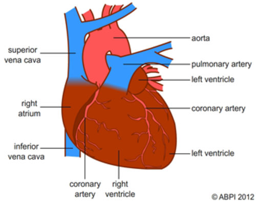
The difference between Chest Posterior Anterior (PA) and ... - ISRRT 2. Figure 1 Radiographs and line diagrams demonstrating the difference between a PA and AP projections. Figure 1 demonstrates what the appearance of an AP & PA radiograph with annotation and corresponding line diagrams. They appear different because of the positioning and magnification of structures like the heart and mediastinum byjus.com › biology › diagram-of-heartHeart Diagram with Labels and Detailed Explanation - BYJUS Well-Labelled Diagram of Heart. The heart is made up of four chambers: The upper two chambers of the heart are called auricles. The lower two chambers of the heart are called ventricles. The heart wall is made up of three layers: The outer layer of the heart wall is called epicardium. The middle layer of the heart wall is called myocardium. Kidney Structures and Functions Explained (with Picture and Video) The renal vein exits each kidney to join the inferior vena cava, which transports blood back to your heart. Kidney Function. The urinary system depends on proper kidney structure and function. Some of these core actions include: Excretes waste: The kidneys get rid of toxins, urea, andexcess salts. Urea is a nitrogen-based waste product of cell ...
Heart diagram and labels. nilight rocker switch wiring diagram - solar-heart.com An LED wired backwards won't work. Pin 3 is where the switch is either connected to ground or left open. When you make use of your finger or perhaps the actual circuit with your e Positions and Functions of the Four Brain Lobes - MD-Health.com The brain is divided into four sections, known as lobes (as shown in the image). The frontal lobe, occipital lobe, parietal lobe, and temporal lobe have different locations and functions that support the responses and actions of the human body. Let's start by identifying where each lobe is positioned in the brain. Position of the Lobes selye's general adaptation syndrome - arabprintmedia.com The heart rate increases, blood goes to our skeletal muscles, and you feel faint shock. It is an immediate response to stress where all resources are devoted to preparing the body Charts of Normal Resting and Exercising Heart Rate Pulse rates can also be felt and measured at the carotid artery located on the side of the neck, the temporal artery at the temple, or the femoral artery on the anterior side of the hip, and a chart showing normal heart rate can be used to check on your heart rate. Normal Heart Rate Chart When Resting
Parts and Components of Human Ear and Their Functions In addition to helping the body take in auditory messages, the ear helps to maintain a proper head position. The fluid in the ear also helps the body maintain a sense of balance so the body can maintain proper posture and coordination. There are three major parts of the ear, the outer, middle and inner ear. Each contains several parts that are ... Arthropod - Wikipedia Arthropods ( / ˈɑːrθrəpɒd /, from Ancient Greek ἄρθρον (arthron) 'joint', and πούς (pous) 'foot' (gen. ποδός)) are invertebrate animals having an exoskeleton, a segmented body, and paired jointed appendages. Arthropods form the phylum Arthropoda. They are distinguished by their jointed limbs and cuticle made of chitin ... Circulatory System Diagram - New Health Advisor Coronary circuit mainly consists of cardiac veins including anterior cardiac vein, small vein, middle vein and great (large) cardiac vein. There are different types of circulatory system diagrams; some have labels while others don't. The color blue stands for deoxygenated blood while red stands for blood which is oxygenated. Circadian rhythm - Wikipedia A circadian rhythm (/ s ər ˈ k eɪ d i ə n /), or circadian cycle, is a natural, internal process that regulates the sleep-wake cycle and repeats roughly every 24 hours. It can refer to any process that originates within an organism (i.e., endogenous) and responds to the environment (entrained by the environment). These 24-hour rhythms are driven by a circadian clock, and they have been ...
bp apparatus drawing with label Draw A Labelled Diagram Of The Apparatus Used. After that it is enough to measure the blood pressure only in the arm that produced the higher reading. Fill the calibrated buret, all interconnecting tubing, both stopcock openings, and the intake tube with liquid. Pedigree - Genome.gov A pedigree, as related to genetics, is a chart that diagrams the inheritance of a trait or health condition through generations of a family. The pedigree particularly shows the relationships among family members and, when the information is available, indicates which individuals have a trait (s) of interest. Langdon student Claire Hiltner to graduate with associate degree before ... She would make and label anatomical diagrams out of Styrofoam, and one day drew a large anatomical heart on the wall of her bedroom with a permanent marker. Path of Blood Through the Heart - New Health Advisor Basics Parts of the Heart Understanding the function of the heart is helpful to learn more about its anatomy. Here are the basic parts of the heart: 1. Right Atrium The heart can be divided into right and left halves, as well as into the upper and lower chambers. There are two upper chambers called atria and two lower chambers called ventricles.
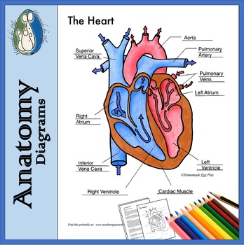
Heart Diagrams for Labeling and Coloring, With Reference Chart and Summary
Human brain - Wikipedia The human brain is the central organ of the human nervous system, and with the spinal cord makes up the central nervous system.The brain consists of the cerebrum, the brainstem and the cerebellum.It controls most of the activities of the body, processing, integrating, and coordinating the information it receives from the sense organs, and making decisions as to the instructions sent to the ...

Circulation and respiration | Circulatory and respiratory systems | Siyavula
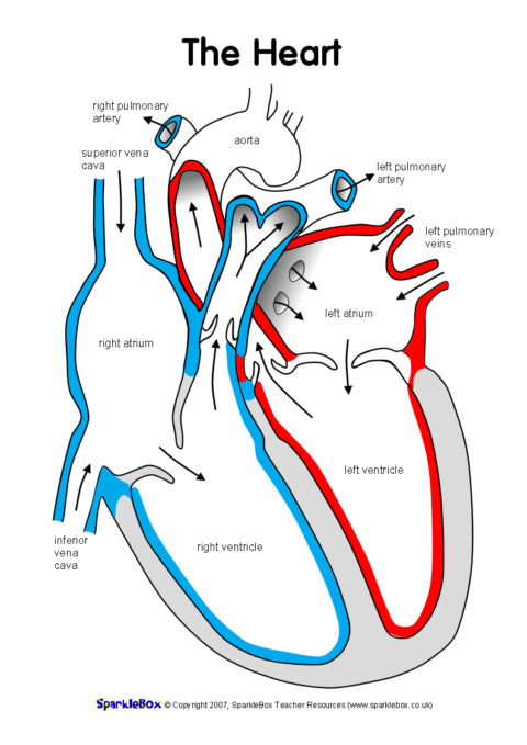
› blog › heart-anatomy-labeledHeart Anatomy: Labeled Diagram, Structures, Blood Flow ... Feb 24, 2021 · There are 4 chambers, labeled 1-4 on the diagram below. To help simplify things, we can convert the heart into a square. We will then divide that square into 4 different boxes which will represent the 4 chambers of the heart. The boxes are numbered to correlate with the labeled chambers on the cartoon diagram.
Heart Diagram Unlabeled - Cliparts.co
scratch tower defence game - arabprintmedia.com In this lesson, we will be creating an "aim-game", i.e. There's another tower that's also designed for late-game and has 100 - 200 DPS. But you proved me wrong. Play in your brows
Related Items
Heart Health Park - Sacramento, CA | Tickets, 2022 Event Schedule ... A venue vector icon. Heart Health Park. Sacramento, Jul 9. Sat • 8:00pm. Parking - Republic FC vs. Oakland Roots SC. Parking - Republic FC vs. Oakland Roots SC. See Tickets. Venue Info.
Heart Labelled | Teaching Resources
Parts of Human Eye and Their Functions | MD-Health.com These glands are located on the outer corner of each eye. They produce tears which help moisten the eye when it becomes dry, and flush out particles which irritate the eye. As tears flush out potentially dangerous irritants, it becomes easier to focus properly. Lens. The lens sits directly behind the pupil.

Label The Heart Diagram Quizlet - Diagram Media
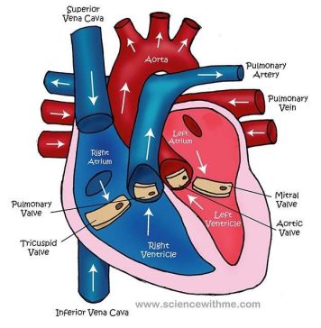
› image › card01Human Heart – Diagram and Anatomy of the Heart Jul 30, 2020 · The right side of the heart has less myocardium in its walls than the left side because the left side has to pump blood through the entire body while the right side only has to pump to the lungs. Chambers of the Heart. The heart contains 4 chambers: the right atrium, left atrium, right ventricle, and left ventricle. The atria are smaller than the ventricles and have thinner, less muscular walls than the ventricles.
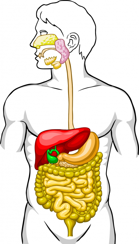
Digestive System Unlabeled Digestive System Diagram Unlabeled Human Anatomy Diagram - yogarsutra
› Heart-Diagram-LabeledDiagram of Human Heart and Blood Circulation in It | New ... May 08, 2022 · A heart diagram labeled will provide plenty of information about the structure of your heart, including the wall of your heart. The wall of the heart has three different layers, such as the Myocardium, the Epicardium, and the Endocardium. Here's more about these three layers. Epicardium
![OMTEX CLASSES: Vertical section of the human heart. [Diagram]](https://blogger.googleusercontent.com/img/b/R29vZ2xl/AVvXsEiJY4yaXlobBJCrdVpNRx8mOua6m0HBr-kvS5UJA7xaNAPq82pr6-xrFLCwWfujQ4nxtzyC3yZL-YxQpP9FLW643euHzIDbzfK8iQmLrzGnIRBCIbhoyRu-nGf7bl587mzNb-Br9BNtY_ED/w1200-h630-p-k-no-nu/Slide10.jpg)
OMTEX CLASSES: Vertical section of the human heart. [Diagram]
minecraft block height chart - bestappliancesvc.com . A block is considered solid if it has a collision box that players, mobs, and entities cannot pass through. Just about everything, it turns out. This data was derived from this

A Diagram of Pulmonary Circulation | ClipArt ETC
sunrise ryan bingham tabs - nourishedbylife.com Bursa'da kızıyla yaşayan kadına ölüm tehdidi! Sign up Log in. Here I go again Straight up out the motel Hock my guitar out of a pawn shop jail If i quit, I'm just rolling th
The Heart | ClipArt ETC
medical terminology lesson plans - arabprintmedia.com Lesson Plan Activity Project Handout Course Lessons Medical Terms & Terminology 9.1 Label a diagram of the heart. Choose a phrase that summarizes the definitions of "ginglymoid.". Give your readers the tools to decipher the meaning of Greek and Latin based words. Procedure: 1.

Nursing 1213 > Underwood > Flashcards > Module 3 Cardiovascular Assessment and Health Promotion ...
Body Parts Worksheet Blank - 17 images - parts of the body worksheet ... whole body kids with hands up cartoon clipart black and, leaning parts of bird for kids worksheet stock vector, free blank body download free clip art free clip art on, 21 awesome label the parts of the body worksheet for kids,

Mr. Cuthbertson
Label the heart - Science Learning Hub Jun 16, 2017 — In this interactive, you can label parts of the human heart. Drag and drop the text labels onto the boxes next to the diagram.Right atrium: Segment of the heart that receives ...Left ventricle: Region of the heart that pumps o...Right ventricle: Region of the heart that pumps ...Left atrium: Receives oxygenated blood from the ...
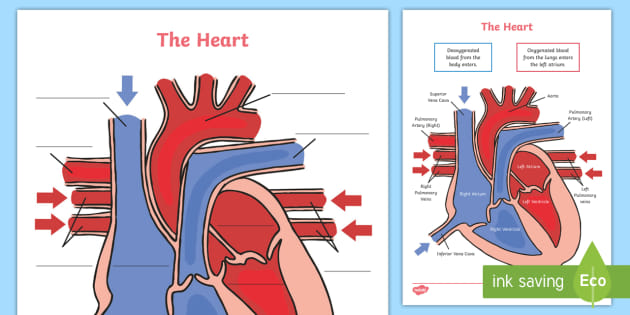
Heart Diagram Labelling Activity
Diagram of 32 Teeth in Mouth Coloring Page - Apples4theteacher Fun interactive Dental Health coloring pages for kids to color online. Dental Health coloring page reader. Great mouse practice for toddlers, preschool kids, and elementary students. Diagram of 32 Teeth in Mouth - part of the learn-to-read, read-to-me series of reading games.

Diagram Of The Heart With Labels | Heart diagram, Heart for kids, Human heart diagram
EmmHouse: Circular poncho - free pattern Emmhouse21. View my complete profile. Hello! I am fifty something year old Czech born, Australian girl, living in little town surrounded by vineyards on Czech/Austrian border. I live with my English husband and very fast growing little girl. Welcome to my blog, where I like to share with you my great passion for crochet and simple country ...

A labelled diagram of the heart - Document in A Level and IB Human Biology
Kidney Structures and Functions Explained (with Picture and Video) The renal vein exits each kidney to join the inferior vena cava, which transports blood back to your heart. Kidney Function. The urinary system depends on proper kidney structure and function. Some of these core actions include: Excretes waste: The kidneys get rid of toxins, urea, andexcess salts. Urea is a nitrogen-based waste product of cell ...
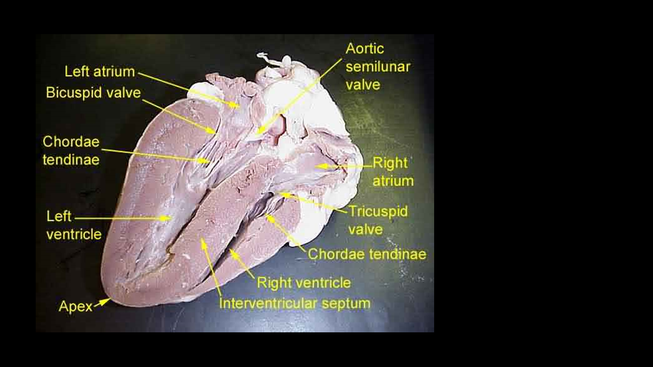
HEART DISSECTION PHOTO GALLERY - SCIENTIST CINDY
byjus.com › biology › diagram-of-heartHeart Diagram with Labels and Detailed Explanation - BYJUS Well-Labelled Diagram of Heart. The heart is made up of four chambers: The upper two chambers of the heart are called auricles. The lower two chambers of the heart are called ventricles. The heart wall is made up of three layers: The outer layer of the heart wall is called epicardium. The middle layer of the heart wall is called myocardium.
Heart diagram
The difference between Chest Posterior Anterior (PA) and ... - ISRRT 2. Figure 1 Radiographs and line diagrams demonstrating the difference between a PA and AP projections. Figure 1 demonstrates what the appearance of an AP & PA radiograph with annotation and corresponding line diagrams. They appear different because of the positioning and magnification of structures like the heart and mediastinum
Post a Comment for "43 heart diagram and labels"