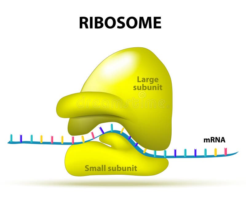
43 ribosome diagram with labels
Ribosomes vector illustration - VectorMine Most Vector Editing Software. 3. High-resolution JPG image. 3800 x 3965 px. License terms in short: Use for everything except reselling item itself. Read a full license here. Description: Ribosomes vector illustration. Anatomical and medical labeled scheme with tRNA, Amino acid, protein, cell, small and large subunit, mRNA. Ribosomes - Definition, Structure, Size, Location and ... Ribosomes are made of proteins and ribonucleic acid (abbreviated as RNA), in almost equal amounts. It comprises of two sections, known as subunits. The tinier ...
DNA Labeling: Transciption and Translation Label the diagram. 1. _____ 5. ... How does the ribosome know the sequence of amino acids to build? 12. What is the difference between a codon and an anticodon? 13. Where does transcription take place? Where does translation take place? 14. Summarize the relationship between proteins and genes.

Ribosome diagram with labels
Plant Cell Diagram Ribosome Functions May 05, 2021 · Ribosomes are a type of organelle. Ribosomes are small organelles of a cell having a dense feature and helps in protein fabrication. They are situated in the cytosol, some bound and free-floating to the membrane of the coarse endoplasmic reticulum. The ribosomes' structure is the same in all cells but smaller in prokaryotic cells. Animal Cell Diagram with Label and Explanation: Cell ... Ribosomes. These are the smallest organelles of the cell comprising 60% RNA and 40% protein; these are also known as RNA-rich sites and are responsible for protein synthesis. Mitochondrion. These are the rod-shaped organs in the cytoplasm which are known as powerhouses of cells that convert oxygen into energy. Endoplasmic Reticulum Cell Organelles- Definition, Structure, Functions, Diagram In the case of prokaryotic cells, the ribosomes are of the 70S with the larger subunit of 50S and the smaller one of 30S. Eukaryotic cells have 80S ribosomes with 60S larger subunit and 40S smaller subunit. Ribosomes are short-lived as after the protein synthesis, the subunits split up and can be either reused or remain broken up.
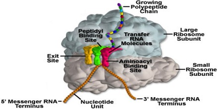
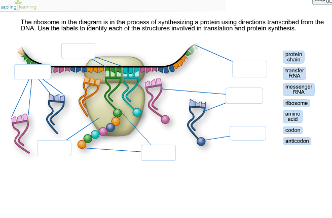
Ribosome diagram with labels. Ribosome and protein synthesis, diagram - Stock Image ... Ribosome and protein synthesis, diagram. C029/3019. Rights Managed. 50.0 MB (867.0 KB compressed) 4827 x 3620 pixels. 40.9 x 30.7 cm ⏐ 16.1 x 12.1 in (300dpi) This image is not available for purchase in your country. Please contact your Account Manager if you have any query. Request Price Add To Basket. Ribosome - Wikipedia Prokaryotic ribosomes are around 20 nm (200 Å) in diameter and are composed of 65% rRNA and 35% ribosomal proteins. Eukaryotic ribosomes are between 25 and 30 nm (250-300 Å) in diameter with an rRNA-to-protein ratio that is close to 1. Solved The ribosome in the diagram is in the process of ... The ribosome in the diagram is in the process of synthesizing a protein using directions transcribed from the DNA. Use the labels to identify each of the structures involved in translation and protein synthesis. Question: The ribosome in the diagram is in the process of synthesizing a protein using directions transcribed from the DNA. Animal Cells: Labelled Diagram, Definitions, and Structure Ribosomal RNA or rRNA combines with proteins to form the basic units of ribosomes. When the units are done, the nucleus spits them out of the nuclear envelope, where they are assembled into ribosomes. The nucleus sends orders in the form of messenger RNA, or mRNA. The messages are sent to ribosomes, which carry out the orders in the rest of the ...
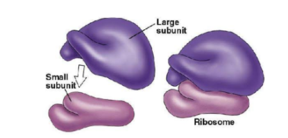
Labeled Plant Cell With Diagrams - Science Trends The ribosomes are created in the nucleolus of the cell. Ribosomes are made out of two smaller subunits, a large ribosomes subunit and a small ribosomal subunits. The transfer RNA or tRNA encodes the correct series of genetic instructions into the mRNA or messenger RNA, which is what ensures that the right proteins are created. Structure of Ribosome (With Diagram) - Biology Discussion A bacterial ribosome is about 250 nm in diameter and consists of two subunits, one large and one small. Both subunits consist of one or more molecules of rRNA and an array of ribosomal proteins. ADVERTISEMENTS: Association of two subunits is called mono-some. The structure of prokaryotic ribosome is given in the figure 8.2 B. Ribosomes: Structure, Composition, and Assembly (With Diagram) Ribosomes in the cytoplasm of eukaryotic cells have a sedimentation coefficient of about 80 S (MW about 4.5 x 10 6) and are composed of 40 S and 60 S subunits. In prokaryotic cells, ribosomes are typically about 70 S (MW about 2.7 x 10 6) and are formed from 30 S and 50 S subunits. Ribosomes Stock Illustrations - 638 Ribosomes Stock ... 638 ribosomes illustrations & vectors are available royalty-free. Reset All Filters. Ribosomes. Vector illustration of ribosomes, RNA and polypeptide chain. Function of ribosomes. Cell with organelles: nucleus, mrna, proteins, tRNA and Ribosome. Process of translating mRNA into protein. vector for medical. Ribosomes vector illustration.
Protein Synthesis Labeling.pdf - 1. Label the diagram ... Protein DNA Amino Acid mRNA Codon tRNA Ribosome Anticodon 2. Label the diagram. Protein Amino Acid Large subunit - rRNA tRNA mRNA codon small subunit rRNA 3. What is the role of mRNA in the process of protein synthesis? The role of mRNA in protein synthesis is that it carries copies of genetic instructions (that tell the cell how to assemble ... What Are Ribosomes? - Definition, Structure and its Functions Ribosomes are located inside the cytosol found in the plant cell and animal cell. The ribosome structure includes the following: It is located in two areas of cytoplasm. Scattered in the cytoplasm. Prokaryotes have 70S ribosomes while eukaryotes have 80S ribosomes. Around 62% of ribosomes are comprised of RNA, while the rest is proteins. Ribosome and protein synthesis, diagram - Stock Image ... Diagram showing protein synthesis in cells (translation). Messenger ribonucleic acid (mRNA, blue with coloured nucleotides) is read by a ribosome (pink). The molecules of transfer RNA (tRNA, key-shaped) each bring an amino acid (orange dot) to bind to the ribosome's protein synthesis site. Structure of Ribosome - Biology Wise Diameter of Ribosome is 20nm. Their composition can be divided into two parts – 2/3 part of r-RNA (ribosomal RNA) and 1/3 part RNP (Ribosomal protein or Ribonuclep protein). Polypeptide chain is fabricated by translating mRNA (messenger RNA) with the aid amino acids that tRNA (transfer RNA) delivers.
About Ribosome - Assignment Point
Bio 1113 - Unit 11 - Gene Expression Flashcards | Quizlet In the following diagram of a ribosome, assign the correct labels: Label 1: a tRNA attached to a polypeptide is found in this area of the ribosome Label 2: a tRNA attached to a single amino acid enters here Label 3: a tRNA that is not attached to anything exits here Label 4: a tRNA molecule Label 5: growing polypeptide Label 6: mRNA being ...
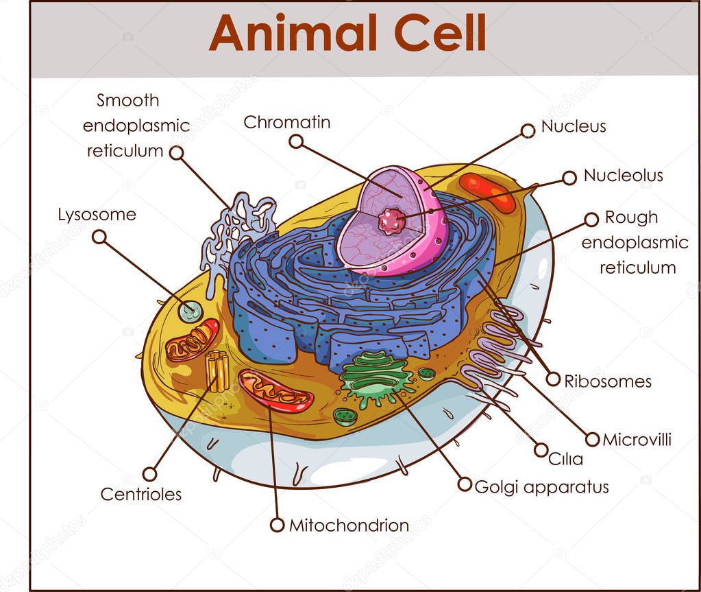
Picture: diagram of a animal cell | Animal Cell Anatomy Diagram Structure with all parts nucleus ...
PDF Quick Review Transcription and Translation label the diagram. 2. ... 910dnamrnait carries the genetic code from dna to ribosome to make a proteinit carries the amino acids to make proteinbecause the genetic code is the recipe to make a protein and is contained in a mrnacodons are in mrna and anti codons are groups of 3 bases in trnatranscription takes place in nucleus; translation takes ...

Ribosome and protein synthesis, diagram - Stock Image - C029/3020 - Science Photo Library
protein synthesis diagram labeled - TheFitnessManual protein synthesis diagram labeled May 8, 2021 by Marie June Table of Contents 1. Transcription units the stage for Translation 2. Making protein is the aim of translation 3. Protein Chemistry Evaluation Quiz 4. Messenger RNA (mRNA) codes for proteins 5. Switch RNAs (tRNAs) deliver amino acids to the ribosome. - "protein synthesis diagram labeled"
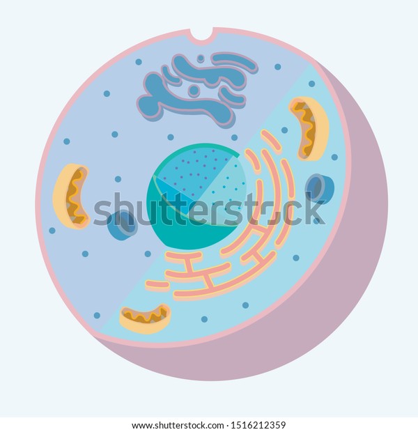
Diagram Animal Cell Organelle Without Labels Stock Vector (Royalty Free) 1516212359
Nucleus and ribosomes (article) | Khan Academy Diagram of the parts of the nucleus of a eukaryotic cell. Image credit: OpenStax Biology. How do ...

8 best Human Physiology images on Pinterest
Animal Cell Diagram - Science Trends An animal cell diagram is a great way to learn and understand the many functions of an animal cell. The diagram, like the one above, will include labels of the major parts of an animal cell including the cell membrane, nucleus, ribosomes, mitochondria, vesicles, and cytosol.

Protein synthesis biological vector illustration scientific diagram | Protein biology, Teaching ...
Solved In the following diagram of a ribosome, assign the ... in the following diagram of a ribosome, assign the correct labels. 5' end of the mrna growing polypeptide a trna attached to a single amino acid ontors here large subunit atrna attached to a polypeptide is found in this area of the nibosome a trna that is not attached to anything exits hore 3' end of the mrna a trna moleculo mossenger rna being …

In The Following Diagram Of A Ribosome Assign The Correct Labels - Free Diagram For Student
Label Transcription and Translation - 7355418.pdf - 1 ... View Label Transcription and Translation - 7355418.pdf from BIOLOGY 101 at Harmony School of Innovation Fort Worth. 1. Label the diagram. DNA Protein Amino Acid Ribosome mRNA Codon tRNA Anticodon 2.

Biology dictionary ribosome - essaycorrections.web.fc2.com
A Labelled Diagram Of Mitochondria with Detailed Explanation Mitochondria are a double-membrane-bound cell organelle found in most eukaryotic organisms. In all living cells, these cell organelles are found freely floating within the cytoplasm of the cell. The diagram of Mitochondria is useful for both Class 10 and 12. It is one among the few topics having the highest weightage of marks and is majorly ...
Write a short note on the ribosome class 9 biology CBSE
Site-specific labeling of the ribosome for single-molecule ... by M Dorywalska · 2005 · Cited by 149 — (C) Time plot of delivery of EF-Tu/GTP/Phe-tRNAPhe (Cy5-acp3U47) to Cy3-labeled ribosomal complexes as detected by an increase in the number of particles ...

Ribosome Structure - Biology for Everybody
Ribosome - Definition, Function and Structure | Biology ... A. Ribosomes translate the 4 base language of DNA into the 20 base language of proteins, allowing for many more combinations. B. The 4 different nucleobases of DNA can be recombined endlessly to produce new proteins. C. Ribosomes can modify proteins with carbohydrates to make them unique. Answer to Question #2 3.

In The Following Diagram Of A Ribosome Assign The Correct Labels - Wiring Site Resource
Biology: parts of a ribosome Diagram | Quizlet sequence of THREE nucleotides that codes for one amino acid mRNA messenger RNA; large family of RNA molecules that convey genetic information from DNA to the ribosomes. large subunit attaches to the small subunit, has three sites E, P, and A small subunit site of mRNA binding E site where tRNA can exit ribosome P site

Chapter 3 Organelles & Cell Membrane - R.E.C.H.S. Biology
Blood Histology Slides with Description and Labeled Diagram The blood is a specialized connective tissue that is fluid and circulates through the vascular channel. In the blood histology slide, you will find different types of cells with their specific features. This might be a short article where I will show you all the cells from the blood microscope slide with a labeled diagram and actual pictures.
Ribosome stock vector. Illustration of science, occurs - 54691349
Ribosome - protein factory - definition, function ... The protein translation by a ribosome consists of three stages: (1) Initiation, (2) Elongation, and (3) Termination. Initiation - the ribosome assembles around the target mRNA. A small ribosome subunit links onto the "start-end" of an mRNA strand. "Initiator tRNA" also enters the small subunit and binds to the start codon (most commonly, AUG).
Solved: The Ribosome In The Diagram Is In The Process Of S... | Chegg.com
Ribosomes- Definition, Structure, Functions and Diagram Apr 02, 2021 · Ribosomes Definition The ribosome word is derived – ‘ribo’ from ribonucleic acid and ‘somes’ from the Greek word ‘soma’ which means ‘body’. Ribosomes are tiny spheroidal dense particles (of 150 to 200 A0 diameters) that are primarily found in most prokaryotic and eukaryotic. They are sites of protein synthesis.
Translation
Cell Organelles- Definition, Structure, Functions, Diagram In the case of prokaryotic cells, the ribosomes are of the 70S with the larger subunit of 50S and the smaller one of 30S. Eukaryotic cells have 80S ribosomes with 60S larger subunit and 40S smaller subunit. Ribosomes are short-lived as after the protein synthesis, the subunits split up and can be either reused or remain broken up.

Copy of "Plant cell"
Animal Cell Diagram with Label and Explanation: Cell ... Ribosomes. These are the smallest organelles of the cell comprising 60% RNA and 40% protein; these are also known as RNA-rich sites and are responsible for protein synthesis. Mitochondrion. These are the rod-shaped organs in the cytoplasm which are known as powerhouses of cells that convert oxygen into energy. Endoplasmic Reticulum

Ribosomi - Struttura, Funzione e Sintesi Proteica - BioPills
Plant Cell Diagram Ribosome Functions May 05, 2021 · Ribosomes are a type of organelle. Ribosomes are small organelles of a cell having a dense feature and helps in protein fabrication. They are situated in the cytosol, some bound and free-floating to the membrane of the coarse endoplasmic reticulum. The ribosomes' structure is the same in all cells but smaller in prokaryotic cells.

Structure of Ribosome | Cell Organelles | Pinterest | Teaching biology, Ap biology and Molecular ...
Post a Comment for "43 ribosome diagram with labels"