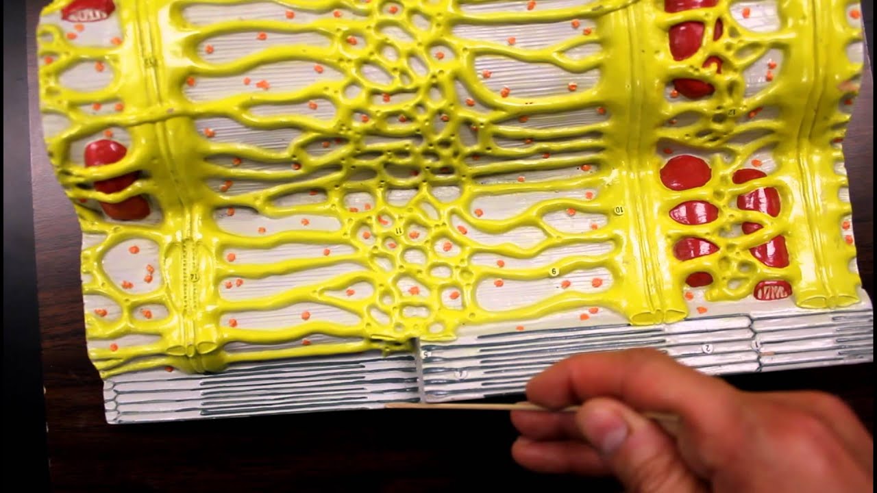
41 muscle fiber model with labels
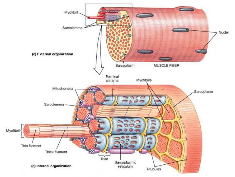
10.2 Skeletal Muscle - Anatomy & Physiology Figure 10.2.2 - Muscle Fiber: A skeletal muscle fiber is surrounded by a plasma membrane called the sarcolemma, which contains sarcoplasm, the cytoplasm of muscle cells. A muscle fiber is composed of many myofibrils, which contain sarcomeres with light and dark regions that give the cell its striated appearance. The Sarcomere Muscular System Labeled Diagram Pictures, Images and Stock Photos Labeled Muscles of the Human Body, Anterior View, 3D Rendering. Frontal view of the muscular system of the male human body with descriptive labels pointing to the muscles on a white background. Diagram of Front Muscles of Human Body. Tendons and Ligaments in Hand and Foot. Female Anterior Leg Muscles Labeled on White.
Altay Size: 110x19x14,5 Weight: approx. 5100 g. Full Description. Muscular Body - cod:6000.58. This 1/4 life-size model is a useful tool to study human superficial musculature. Significant structures are numbered and referenced on the accompanying k-card. Size: 25x18x40 cm Weight: 765 g. Full Description. Skeletal Muscle Fiber - cod:6000.32. This ...

Muscle fiber model with labels
Sarcomere (Muscle) Coloring | Human anatomy and physiology ... - Pinterest Learn the structure of a muscle fiber by coloring an individual sarcomere. Myofilaments, mitochondria, annd tubules can all be identified and labeled on this image. Biologycorner 17k followers More information Muscles are composed of fibers, this coloring exercise asks you to identify the individual parts of the sarcomere. Learn all muscles with quizzes and labeled diagrams | Kenhub Human body muscle diagrams. Muscle diagrams are a great way to get an overview of all of the muscles within a body region. Studying these is an ideal first step before moving onto the more advanced practices of muscle labeling and quizzes. If you're looking for a speedy way to learn muscle anatomy, look no further than our anatomy crash courses . Anatomy Model Keys - Anatomical Models and Keys - NEOMED Library at ... Northeast Ohio Medical University is an Equal Education and Employment Institution ADA Compliance | Title IX. NEOMED Library- 4209 St, OH-44, Rootstown, OH 44272 - "A Building" Second Floor. 330-325-6600. library@neomed.edu. Except where otherwise noted, content on the NEOMED LibGuides is licensed for reuse under a Creative Commons 3.0 Attribution-NonCommercial license (CC BY-NC)
Muscle fiber model with labels. Muscle Anatomy Models Anatomy Now is the leading provider of 3D human anatomical muscle system models, charts, and replica models for the medical profession and education of patients. There are three distinct types of muscles: skeletal muscles, cardiac or heart muscles, and smooth muscles. Skeletal Muscle Fiber | Types, Characteristics & Anatomy - Video ... These differences in muscle capability are the result of different types of skeletal muscle fibers. These types are: Type 1 muscle fibers. Type 2A muscle fibers. Type 2B muscle fibers. Type 1 ... Muscle Fibers: Anatomy, Function, and More - Healthline Muscle fibers are single muscle cells. When grouped together, they work to generate movement of your body and internal organs. You have three types of muscle tissue: skeletal, smooth, and cardiac.... Muscle Models | Muscle Figures | Musculature Models 3B MICROanatomy™ Human Muscle Fiber Model, 10,000 times magnified - 3B Smart Anatomy $ 339.00 Item: 1000213 [B60] This micro-anatomy model magnifies the anatomy of the human muscle fiber approximately 10,000 times. This muscle model illustrates a section of a skeletal muscle fiber and its neuromuscular end plate.
Skeletal Muscle Fiber Model - Myofibrils - YouTube This video was produced to help students of human anatomy at Modesto Junior College study our anatomical models. Muscles Labeling - The Biology Corner The activity linked below is a drag and drop activity for students to practice labeling the muscles, there are 6 slides showing images of muscles and fibers and the connective tissue surrounding the fibers (endomysium, perimysium, epimysium). Google Slides Key (TpT) Prev Article Next Article muscle fiber model labeling Diagram - Quizlet muscle fiber model labeling STUDY Learn Flashcards Write Spell Test PLAY Match Gravity Created by crlavenuePLUS Terms in this set (7) transverse tubule sarcoplasmic reticulum triad sarcolemma myofibril consists of actin (thin) & myosin (thick) fibrils sarcomere nucleus Subjects Arts and Humanities Languages Math Science Social Science Other Labeled Sarcomere Diagram The thin filaments Look at the diagram above and realize what happens as a muscle contracts. As will soon be described, the functional unit of a skeletal muscle fiber is the sarcomere, a highly organized arrangement of the contractile myofilaments actin .Play this quiz called Label the Sarcomere and show off your skills.
Muscle fiber model Quiz - PurposeGames.com About this Quiz This is an online quiz called Muscle fiber model There is a printable worksheet available for download here so you can take the quiz with pen and paper. Your Skills & Rank Total Points 0 Get started! Today's Rank -- 0 Today 's Points One of us! Game Points 13 You need to get 100% to score the 13 points available Muscle Fiber Model (Altay) Flashcards - Quizlet Muscle fiber model identifications Terms in this set (21) sarcolemma Identify the membrane. endomysium Identify the tissue layer. myofibril Identify the structure. thick myofilament Identify the structure. thin myofilament Identify the structure. neuromuscular junction Identify the connection. axon Identify the structure. axon terminals Muscle Fiber. 1. Myofibrils 2. Mitochondrium 3. Postsynaptic membrane 4 ... Muscle Fiber. 1. Myofibrils 2. Mitochondrium 3. Postsynaptic membrane 4. Synaptic gap with basal lamina 5. Presynaptic membrane 6. Presynaptic vesicle 7. Schwann cell 8. Nucleus 9. Actin filament 10. Sarcomere 11. Myosin filament 12. Myelin sheath 13. Neurofibers 14. Cell membrane (sarcolemma) 15. Transverse membrane tube 16. Triad 17. muscle anatomy labled skeleton human labeled bones diagram anatomy body chart system labels parts diagrams. Gentry, Teresa M / Anatomy Diagrams ... Muscle Fiber With Neuromuscular Junction Model ... muscle fiber with neuromuscular junction model. Muscles muscle cardiac diseases body labeled heart operation ap posted. Aap muscular wiley gross anatomy quiz lab system ...
Muscle Fiber Types: Anatomy and Physiology - YouTube
Muscle Fiber Model #1 - Ohio University - YouTube Muscle Fiber Model #1 - Ohio University - Anatomy & Physiology

muscle fiber diagram | Muscle Fiber: Cell & myofibril | Anatomy & Physiology Stuff | Pinterest ...
Skeletal Muscle Fiber Location and Composition - GetBodySmart A review of skeletal muscle fiber (cell) location, structure, anatomy, and function using interactive animations, models, and labeled diagrams. Start learning now!

Print muscle fibers flashcards | Easy Notecards
PDF Anatomy & Physiology - TMCC Subcutis (Hypodermis) 1. External Horny Layer (Stratum corneum) 1a. Clear Layer (Stratum lucidum) -(KS 3 only) 2. Internal Hornless Germinative Zone (Stratum germinativum) 2a. Granular Layer (Stratum granulosum) 2b. Prickle-cell Layer (Stratum spinosum) 2c. Cylindrical Layer (Stratum basale) 3. Papillae 4.

V Ling: 08.10
Muscle Fiber Labeling Quiz - PurposeGames.com This is an online quiz called Muscle Fiber Labeling Quiz There is a printable worksheet available for download here so you can take the quiz with pen and paper. Your Skills & Rank Total Points 0 Get started! Today's Rank -- 0 Today 's Points One of us! Game Points 17 You need to get 100% to score the 17 points available Actions 2 favs

Neurolemmocyte On Skeletal Muscle Model - Human Anatomy - GUWS Medical
SAC A&P Model Key - Muscular System Muscular System. M1 - Muscled Arm. M2 - Muscle Leg. M3 - Female Muscle Figure. M4 - Microanatomy Muscle Fiber. M5 - Muscle Figure.

Skeletal muscle fiber model
General Anatomy of Skeletal Muscle Fibers - GetBodySmart Skeletal Muscle Fiber Location and Arrangement. are located inside muscles, where they are organized into bundles called […] Internal Anatomy of Skeletal Muscle Fibers. An interactive quiz about the internal anatomy of skeletal muscle fibers, featuring illustrations-based multiple choice questions.
MUSCULAR SYSTEM ANATOMY:Muscle fiber with sarcomere model description - YouTube
Muscular System - Muscles of the Human Body - Innerbody Most of the muscle fiber's structure is made up of myofibrils, which are the contractile structures of the cell. Myofibrils are made up of many proteins fibers arranged into repeating subunits called sarcomeres. The sarcomere is the functional unit of muscle fibers. (See Macronutrients for more information about the roles of sugars and proteins.)
multi choice chapter 10. Muscle Tissue Flashcards | Easy Notecards
Skeletal Muscle | Anatomy and Physiology | | Course Hero Because skeletal muscle cells are long and cylindrical, they are commonly referred to as muscle fibers. Skeletal muscle fibers can be quite large for human cells, with diameters up to 100 μm and lengths up to 30 cm (7.6 in) in the Sartorius of the upper leg.During early development, embryonic myoblasts, each with its own nucleus, fuse with up to hundreds of other myoblasts to form the ...
30 Drag The Labels Onto The Diagram To Identify Structural Features Associated With Skeletal ...
Muscle Model - an overview | ScienceDirect Topics The muscle models have been used in the exoskeleton control schemes. Unlike the dynamic model, the muscle model predicts the muscle forces deployed by the muscles of the human limb joint as a function of muscle neural activities and the joint kinematics (Anam and Al-Jumaily, 2012). The input is the EMG signals and the output is force estimation.

Basics of Muscle Fiber Types - YouTube
anatomy labeled muscle fiber Muscle Cells - Types Of Cells In The Body jatypesofcells.weebly.com. muscle cell diagram cells parts. Sarcolemma lookfordiagnosis.com. sarcolemma improve help muscle fiber. Anatomy muscles posterior human chart muscle medical lithograph frame 1930s drawing structure revisit later favorites sold. Muscle cells.

muscle fibers Archives - Chape Fitness
Anatomy Model Keys - Anatomical Models and Keys - NEOMED Library at ... Northeast Ohio Medical University is an Equal Education and Employment Institution ADA Compliance | Title IX. NEOMED Library- 4209 St, OH-44, Rootstown, OH 44272 - "A Building" Second Floor. 330-325-6600. library@neomed.edu. Except where otherwise noted, content on the NEOMED LibGuides is licensed for reuse under a Creative Commons 3.0 Attribution-NonCommercial license (CC BY-NC)

Anvil Auto by Curtiss Lichty at Coroflot.com
Learn all muscles with quizzes and labeled diagrams | Kenhub Human body muscle diagrams. Muscle diagrams are a great way to get an overview of all of the muscles within a body region. Studying these is an ideal first step before moving onto the more advanced practices of muscle labeling and quizzes. If you're looking for a speedy way to learn muscle anatomy, look no further than our anatomy crash courses .
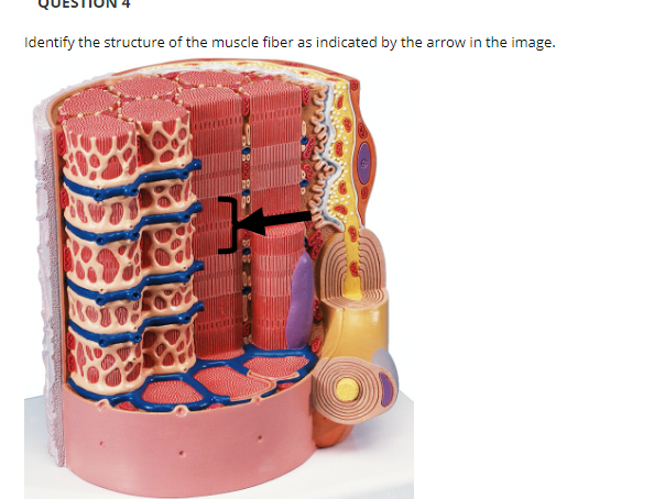
Answered: Identify the structure of the muscle… | bartleby
Sarcomere (Muscle) Coloring | Human anatomy and physiology ... - Pinterest Learn the structure of a muscle fiber by coloring an individual sarcomere. Myofilaments, mitochondria, annd tubules can all be identified and labeled on this image. Biologycorner 17k followers More information Muscles are composed of fibers, this coloring exercise asks you to identify the individual parts of the sarcomere.

A.P. Biology Project - Julia Updike: 22. Muscle Fiber - Striated

Biology 156: April 2008
Drag The Labels Onto The Diagram To Identify Structural Features Associated With Skeletal Muscle ...
Post a Comment for "41 muscle fiber model with labels"