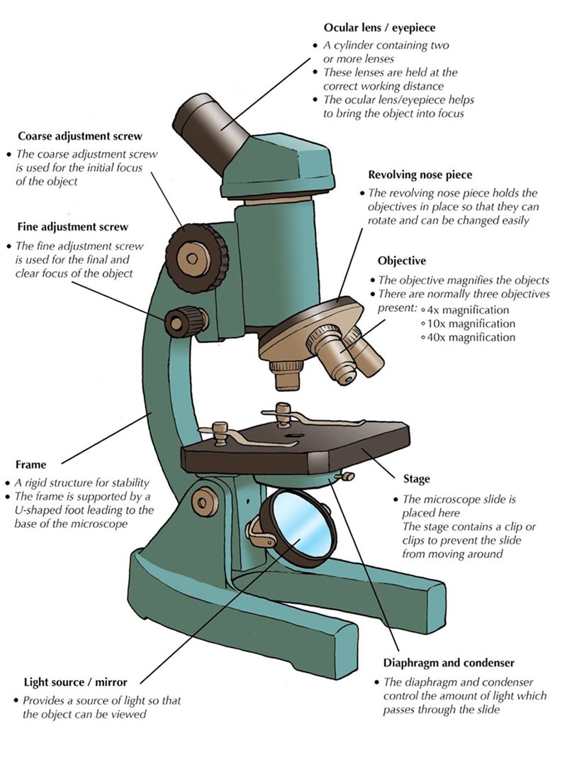
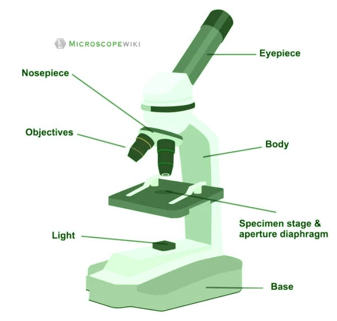
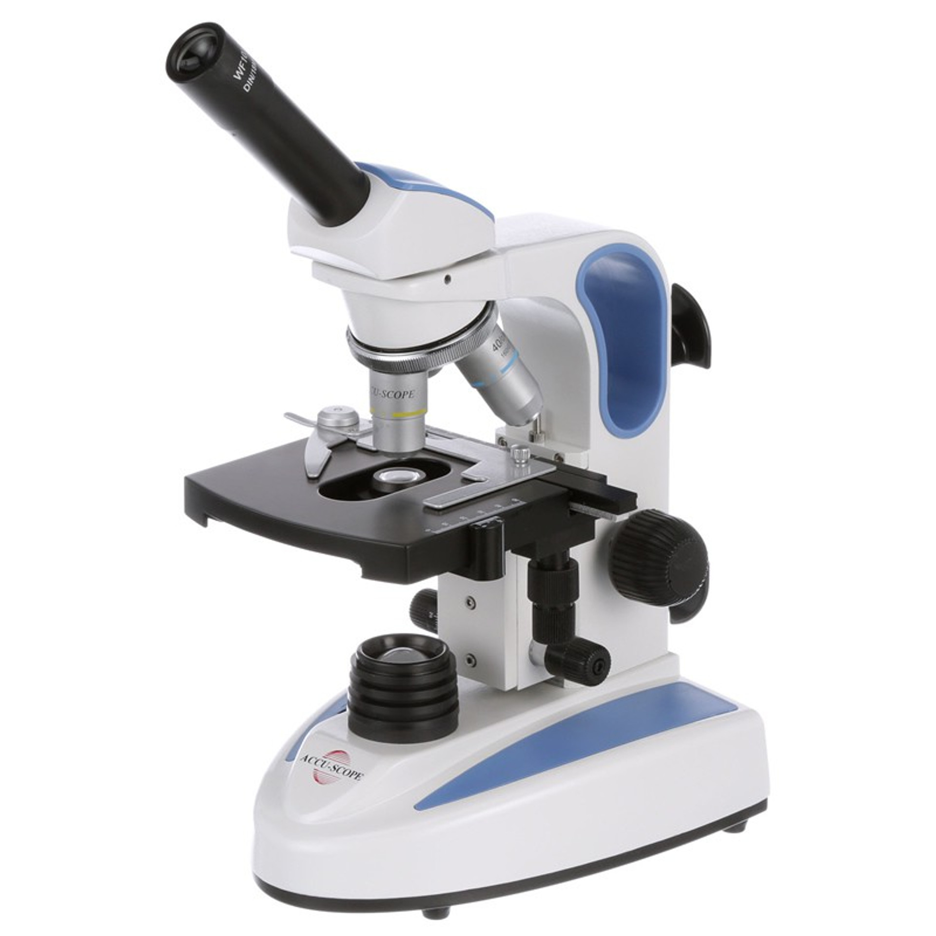
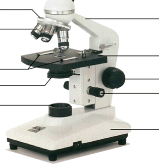
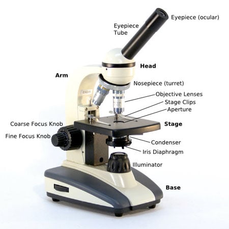
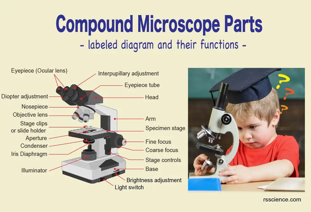
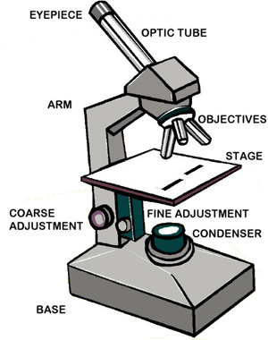
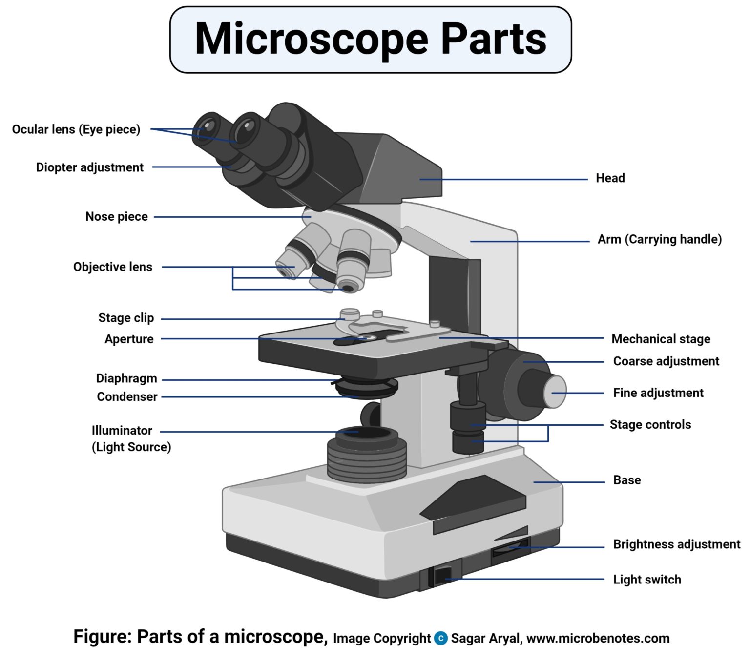
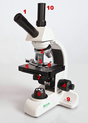
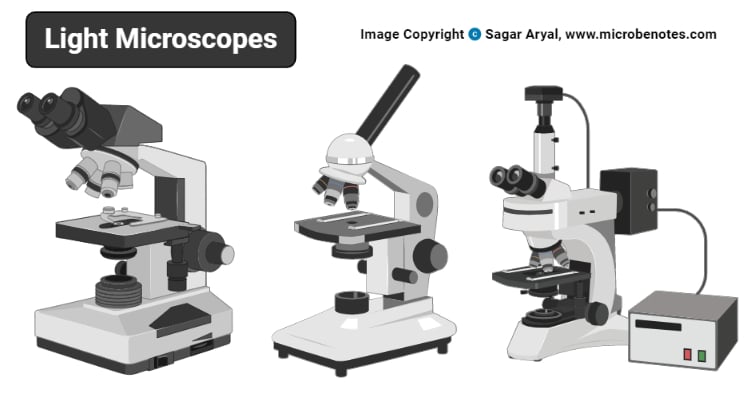
45 easy microscope diagram with labels
Microscope Quiz: How Much You Know About Microscope Parts ... - ProProfs Projects light upwards through the diaphragm, the specimen, and the lenses. 5. Is used to regulates the amount of light on the specimen. Supports the slide being viewed. Moves the stage up and down for focusing. 6. Is used to support the microscope when carried. Moves the stage slightly to sharpen the image. Plant Cell: Diagram, Types and Functions - Embibe Exams These differences can be clearly understood when the cells are examined under an electron microscope. Observe the labelled diagram of plant cell structure as given below: Are Plant Cells Prokaryotic or Eukaryotic? The cell is the basic structural and functional unit of life in all living organisms.
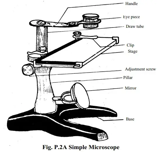
Microscope Types (with labeled diagrams) and Functions Simple microscope labeled diagram Simple microscope functions It is used in industrial applications like: Watchmakers to assemble watches Cloth industry to count the number of threads or fibers in a cloth Jewelers to examine the finer parts of jewelry Miniature artists to examine and build their work Also used to inspect finer details on products

Easy microscope diagram with labels
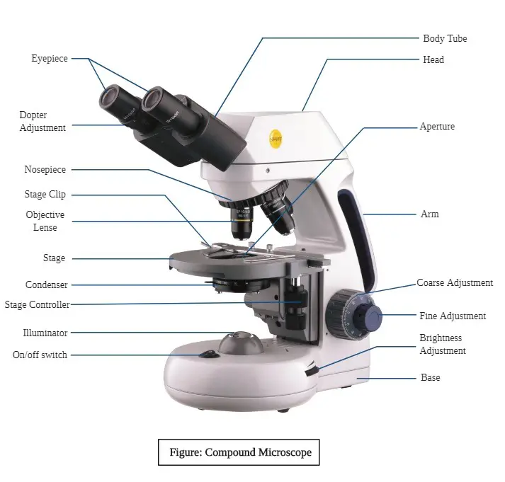
Best indoor home saunas Discover the best portable saunas with easy set-up design. Zipped openings to use devices and a detachable frame for easy storage. ... This indoor sauna kit with 1-9 level to control temperature, easily adjust power and time by a remote control or the button of steamer. ... 【One Box for Whole Family】This is a pretty practical Home Sauna Spa. Home Indoor Saunas 3,686 … Compound Microscope - Diagram (Parts labelled), Principle and Uses See: Labeled Diagram showing differences between compound and simple microscope parts Structural Components The three structural components include 1. Head This is the upper part of the microscope that houses the optical parts 2. Arm This part connects the head with the base and provides stability to the microscope. Colon Histology Slide with Labeled Diagram - AnatomyLearner Now, it will be easy for you to write the identification points of the colon microscope slide. The unique identification points of the colon slide under a light microscope are - The wall (total) of the sample tissue section shows four distinct layers - tunica mucosa, tunica submucosa, tunica muscularis, and tunica serosa layers,
Easy microscope diagram with labels. Simple microscope Class 12, Definition, Magnification, working, Parts ... Definition: A simple microscope is used to view the magnified image of an object. It is made up of a convex lens. The convex lens produces a virtual, erect, and magnified image when the position of the object is within the focal length. Simple Microscope- Definition, Principle, Magnification, Parts ... The magnifying power of a simple microscope is given by: M = 1 + D/F Where D = the least distance of distinct vision F = focal length of the convex lens The focal length of the convex lens should be small because the smaller the focal length of the lens, the greater will be its magnifying power. Microscope: Types of Microscope, Parts, Uses, Diagram - Embibe There are various microscopes, starting from a single lens Simple microscope to a Compound microscope with two lenses; we have various electron microscopes (SEM and TEM) and Light microscopes (confocal). Microscopes have a major role to play when it comes to analyzing tissue or DNA samples or virus strains that help in forensics and biology too. On-chip beam rotators, adiabatic mode converters, and … 07/07/2022 · Though implementing polarization manipulation in free space is easy, arbitrary on-chip polarization control using integrated waveguides remains an open challenge. Existing on-chip polarization ...
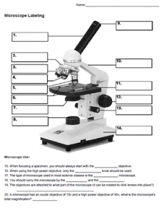
Parts of the Microscope with Labeling (also Free Printouts) Parts of the Microscope with Labeling (also Free Printouts) By Editorial Team March 7, 2022 A microscope is one of the invaluable tools in the laboratory setting. It is used to observe things that cannot be seen by the naked eye. Table of Contents 1. Eyepiece 2. Body tube/Head 3. Turret/Nose piece 4. Objective lenses 5. Knobs (fine and coarse) 6. Inorganic Chemistry 4th edition, Catherine Housecroft Purple acid phosphatases (PAPs) are a group of metallohydrolases that contain a dinuclear Fe(III)M(II) center (M(II) = Fe, Mn, Zn) in the active site and are able to catalyze the hydrolysis of a variety of phosphoric acid esters. Pseudostratified Columnar Epithelium under a Microscope with a Labeled ... The adventitia of a trachea histology slide contains the loose connective tissue with numerous blood vessels. Learn more about trachea histology with a labeled diagram from another article by an anatomy learner. Histological features of the trachea (lining epithelium and other different layers) with a labeled diagram. Microscope Diagram Worksheet - Stock Walker Use the following terms to correctly label the microscope: Use the words from this word list to identify the parts of the microscope. In this worksheet, students will look at the different parts of a light microscope and be able to label one correctly. Microscope Diagram Worksheet - The Microscope Create A Labelled Diagram Teaching Resources ...
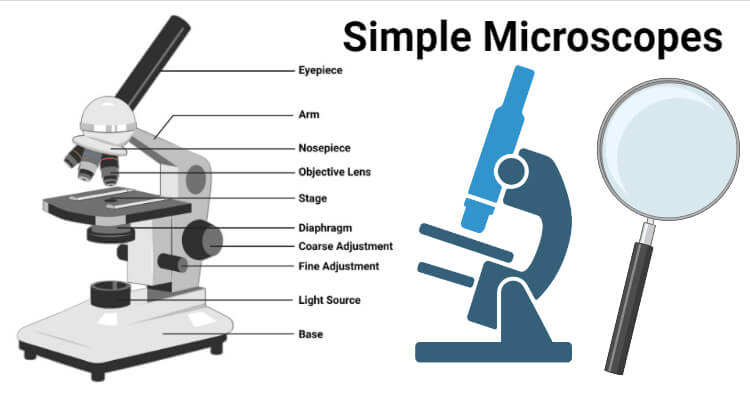
Light Microscope- Definition, Principle, Types, Parts, Labeled Diagram ... Two focusing knobs i.e the fine adjustment knob and the coarse adjustment knob, found on the microscopes' arm, which can move the stage or the nosepiece to focus on the image. the sharpen the image clarity. It has a light illuminator or a mirror found at the base or on the microbes of the nosepiece. Stereo Microscope - Parts, Types and Uses - Laboratoryinfo.com Stereo Microscope Parts and Functions. It has three key parts namely: body, focus block, and viewing head/body. Let us take a look at the functions of every part.. Body/viewing head - It houses the optical parts in the upper section of the microscope. Focus block - It attaches the head of the microscope to the stand and focuses it. Stand - It supports the microscope as well as houses ... Simple Microscope - Diagram (Parts labelled), Principle, Formula and Uses Parts of a Simple Microscope A simple microscope consists of Optical parts Mechanical parts Labeled Diagram of simple microscope parts Optical parts The optical parts of a simple microscope include Lens Mirror Eyepiece Lens A simple microscope uses biconvex lens to magnify the image of a specimen under focus. Cambridge International AS & A Level Biology Coursebook … The light microscope uses light as a source of radiation, while the electron microscope uses electrons, for reasons which are discussed later. y op Microscopes that use light as a source of radiation are called light microscopes. Figure 1.3 shows how the light microscope works. id ie w ge C U R ni ev ve rs w ie 1.3 Plant and animal cells as seen with a light microscope Pr y es …
Simple Squamous Epithelium under a Microscope with a Labeled Diagram ... Simple squamous epithelium under microscope labeled in renal corpuscle The cortex of a kidney consists of renal corpuscles and the convoluted tubule, straight tubules, nephrons, connecting tubules, and collecting ducts. You will find the medullary ray in the medulla of the kidney that comprises straight tubules and collecting ducts.
15 Microscope Parts with Diagram, Location and Function - Study Read A compound microscope has about 15 parts that assist in viewing with a naked eye, a sample holder, a magnifying lens, and a light source. For the convenience of study, we can divide them based on their purpose in the instrument like. A) Parts that assist in viewing the object. B) Part that helps in the adjustment of lenses for a clear view.
Cells Diagram | Science Illustration Solutions - Edrawsoft Cells Diagram. Cells are the basic building blocks of all living things. The human body is composed of trillions of cells. Cells have many parts, each with a different function. Some of these parts, called organelles, are specialized structures that perform certain tasks within the cell. Drawing cells diagram helps you better understand your ...
Jejunum Histology Slide with Labeled Diagram and ... - AnatomyLearner The villus of the jejunum fold lines with the simple columnar epithelium. Within each villus of the jejunum, you will find the lamina propria. ... In the labeled diagram, I tried to show you all these above-mentioned histological features from the jejunum. Normal jejunum wall histology with diagram. ... In the electron microscope, you will find ...
Microscope, Microscope Parts, Labeled Diagram, and Functions Microscope, Microscope Parts, Labeled Diagram, and Functions What is Microscope? A microscope is a laboratory instrument used to examine objects that are too small to be seen by the naked eye. It is derived from Ancient Greek words and composed of mikrós, "small" and skopeîn,"to look" or "see".
Lower Secondary Science LEARNER’S BOOK 8 - Issuu 22/02/2021 · Read Lower Secondary Science LEARNER’S BOOK 8 by Cambridge University Press Education on Issuu and browse thousands of other publications on our p...
ALEX | Alabama Learning Exchange Finally, students will draw a diagram and write an explanation of the apparent movement of stars using data from the graphs and class model. This lesson results from the ALEX Resource Gap Project. View Standards Standard(s): [MA2015] PRE (9-12) 29 : 29 ) (+) Use special triangles to determine geometrically the values of sine, cosine, and tangent for π / 3, π / 4, and π / 6, and …
Electron Microscope- Definition, Principle, Types, Uses, Labeled Diagram Parts of Electron Microscope Electron Microscope is in the form of a tall vacuum column that is vertically mounted. It has the following components: 1. Electron gun The electron gun is a heated tungsten filament, which generates electrons. 2. Electromagnetic lenses The condenser lens focuses the electron beam on the specimen.
Difference between Simple Microscope and Compound Microscope The convex lens, used in a simple microscope, produces a virtual image of the object when it is placed within its focal length. This allows the observers to view the object. The diagram given below shows the structure of a simple microscope with all labeled parts:
K To 12 Science Grade 7 Learners Material - Module Materials Needed food labels Procedure 1. Refer to the labels of different food products below. 36 Soy sauce Ingredients: water, hydrolysed soybean protein, iodized salt, sugar, natural and artificial colors with tartrazine, acidulant, monosodium glutamate, 0.1% potassium sorbate, natural flavor and flavor enhancer.
Parts of Stereo Microscope (Dissecting microscope) – labeled diagram ... They are easy to use (you don’t need to worry about focusing), but at the same time, lack flexibility. Sometimes, you may see a “Dual power” stereo microscope. It means a fixed power stereo microscope with two levels of magnification (commonly, 10x/30x or 20x/40x). Simply rotate the objective housing to click into the desired level of ...
TIR Retroreflector Prisms - Thorlabs 08/07/2020 · After exposure, the optic is examined by a microscope (~100X magnification) for any visible damage. The number of locations that are damaged at a particular power/energy level is recorded. Next, the power/energy is either increased or decreased and the optic is exposed at 10 new locations. This process is repeated until damage is observed. The damage threshold is …
Scanning Electron Microscope (SEM) - Diagram, Working Principle ... Scanning electron microscope is a classification of electron microscope that uses raster scanning to produce the images of a specimen by scanning using a focused electron beam on the surface of the specimen. An SEM creates magnified images of the specimen by probing along a rectangular area of the specimen with a focused electron beam.
Microscopy- History, Classification, Terms, Diagram - The Biology Notes Fluorescence Microscopy is a microscopy technique that uses a fluorescent microscope with a UV light source. It is widely used in detecting antigens, antibodies, and other macromolecules. Fluorescence Microscope 5. Confocal Microscopy Confocal Microscopy is a newer microscopy technique that uses a focused laser beam.
Parts of a microscope with functions and labeled diagram - Microbe Notes Figure: Diagram of parts of a microscope There are three structural parts of the microscope i.e. head, base, and arm. Head - This is also known as the body. It carries the optical parts in the upper part of the microscope. Base - It acts as microscopes support. It also carries microscopic illuminators.
Compound Microscope - Types, Parts, Diagram, Functions and Uses It comes with a wide body and base. Its distinct parts include a condenser, illumination, focus lock, mechanical stage, and a revolving nosepiece which can hold up to five objectives. It usually has a binocular head, which makes long-term observation easy. Image 22: An example of a research compound microscope.
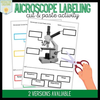
Free Microscope Worksheets for Simple Science Fun for Your Students Parts of a Microscope . The first worksheet labels the different parts of a microscope, including the base, slide holder, and condenser. If you have a microscope, compare and contrast this worksheet to it. Also, your kids can color this microscope diagram in and read the words to each part of the microscope. Then, you can have your children study the words and later have a …
Bright-field microscope (Compound light microscope) - Diagram (Parts ... Bright-field microscope parts (Labeled Diagram) Ocular Lens This microscope has two eye lenses or ocular lens on the top of the microscope that are used to focus the image from the objective lens. It is from these lenses that we see the magnified image of the specimen. Objective Lens
Labeling a Microscope Free Worksheet Pack - Homeschool Giveaways Easy & Free Crafts; ... Once they have the microscope parts labeled, see if they can use the microscope to look at an image of the specimen. ... Put the word bank on the table with a blank diagram of the microscope and see how fast your kids can match up the correct term. You can even print out several of these worksheets and let the kids race ...
Compound Microscope- Definition, Labeled Diagram, Principle, Parts, Uses In order to ascertain the total magnification when viewing an image with a compound light microscope, take the power of the objective lens which is at 4x, 10x or 40x and multiply it by the power of the eyepiece which is typically 10x. Therefore, a 10x eyepiece used with a 40X objective lens will produce a magnification of 400X.
Simple Microscope - Parts, Functions, Diagram and Labelling Simple Microscope - Parts, Functions, Diagram and Labelling By Editorial Team March 7, 2022 A microscope is one of the commonly used equipment in a laboratory setting. A microscope is an optical instrument used to magnify an image of a tiny object; objects that are not visible to the human eyes. Table of Contents
Colon Histology Slide with Labeled Diagram - AnatomyLearner Now, it will be easy for you to write the identification points of the colon microscope slide. The unique identification points of the colon slide under a light microscope are - The wall (total) of the sample tissue section shows four distinct layers - tunica mucosa, tunica submucosa, tunica muscularis, and tunica serosa layers,
Compound Microscope - Diagram (Parts labelled), Principle and Uses See: Labeled Diagram showing differences between compound and simple microscope parts Structural Components The three structural components include 1. Head This is the upper part of the microscope that houses the optical parts 2. Arm This part connects the head with the base and provides stability to the microscope.
Best indoor home saunas Discover the best portable saunas with easy set-up design. Zipped openings to use devices and a detachable frame for easy storage. ... This indoor sauna kit with 1-9 level to control temperature, easily adjust power and time by a remote control or the button of steamer. ... 【One Box for Whole Family】This is a pretty practical Home Sauna Spa. Home Indoor Saunas 3,686 …

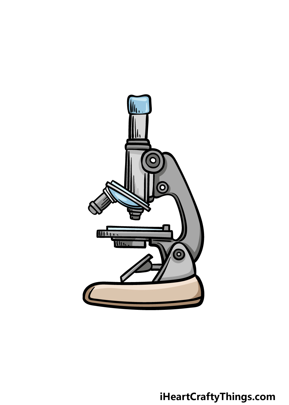
![How To Draw A Microscope Step by Step - [12 Easy Phase]](https://easydrawings.net/wp-content/uploads/2021/01/Overview-for-Microscope-drawing.jpg)













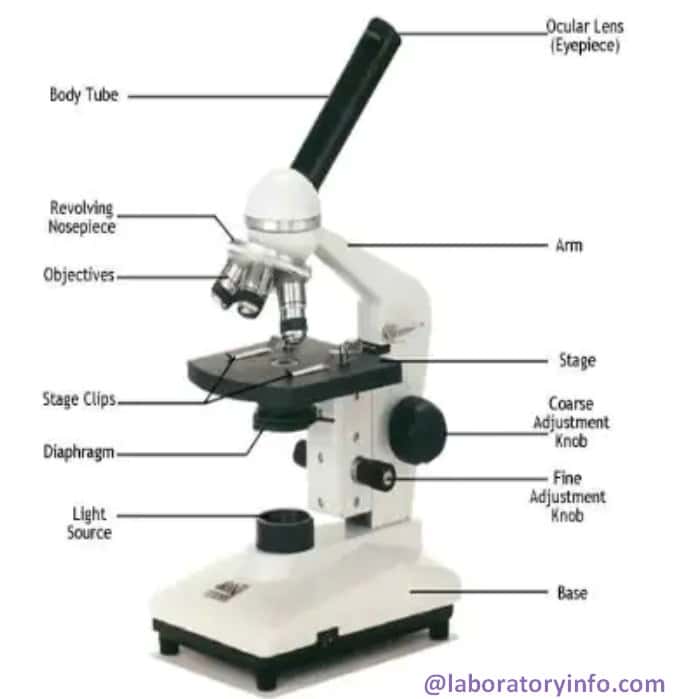


Post a Comment for "45 easy microscope diagram with labels"